Particle-size and shape analysis by means of image analysis
Most particle size measurement methods are based on the assumption of spherical shaped particles. This hypothesis leads to significant errors in the analysis if the particles are flake or rod-shaped. Especially for such highly form-anisotropic particles, image analysis methods provides an excellent alternative for the determination of tailor-made size specifications.
Image analysis methods for the determination of the particle size distribution of a material offers a fundamental advantage over alternative methods such as static light scattering, sedimentation or fractionation (screening): Each particle is photographed individually! This results in several important advantages for the determination of the particle size distribution:
- Realistic proportional values also at the edges of the size distribution, i. e. detection of oversized particles or fine particles
- Visual assessment of the dispersing state of a sample (dispersing quality, agglomerates present)
- Calculation of meaningful size parameters, e. g. geodetic length or Feret diameter for fibres, depending on the application
- Selection of the appropriate distribution type (volume, number) depending on the particular task
In addition, the individual photography of the particles gives the opportunity to make statistical calculations on the particle shape, which in practice enables further differentiation of materials. For example, form anisotropy, the deviation of the particles from the ideal sphere, often plays a decisive role for their application and further processing – for example, the conveyance or compaction of powders, the influence on the rheology in dispersions or, in addition to the particle size distribution, the roughness of the particle surface plays an important role for the success of shaping or polishing.
The necessity for tailor-made particle size and shape parameters, combined with ever-increasing PC processing power, ensures that image analysis methods are becoming increasingly established on the market.
Measurement method
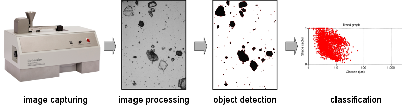
The determination of the particle shape by optical image analysis includes 4 basic steps:
1. Image taking
2. Image processing
3. Object detection
4. Classification
The image taking is ensured by special digital cameras, if necessary in combination to a microscope, to enlarge the particles. The particles may be present neutral (e.g. on an objective) or in motion as well. The dispersing (separation) of particles is possible both in dry-mode (e.g. by simple conveying and riddling or by the usage of compressed air) but also in wet-mode in a solvent. An absolutely basic requirement to perform a successful particle shape analysis is good quality (high resolution and image sharpness, good sample dispersing resulting in individual particles, suitable enlargement etc.). Image processing by appropriate software leads to upgraded pictures: for example isolated pixels and edging particles are eliminated, variations in brightness and signal noise are retouched and connected particles are separated. The main part in object detection is image binarization, whereby every image pixel is assigned to a particle (black) or the background (white) using a threshold. The recognition of objects (particles) and feature attribution is realized by the software. In the last step, the classification, the particles are arranged in classes (e.g. size equivalent classes) on the basis of their attributed features (size and shape parameters).
Numerous size and shape parameters can be determined from the particles images by the appropriate software. Important size parameters are for example (CE) equivalent disc diameter Deq, maximum inscribed disc diameter Din, fibre length XLG (geodesic length) and fibre diameter XFD.
The equivalent disc diameter corresponds to the disc diameter of identical area to 2-D-projected particles, which is often used as size indicator for irregular shaped particles in process technology. On the contrary the maximum inscribed disc diameter of the 2-D-projected particle corresponds more or less to the sieve diameter. The geodesic length and the fibre diameter are suited very well for the characterization of fibres.
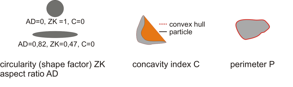
Important size parameters image analysis
There are numerous and very application-specific shape parameters. The aim is to get extra morphological parameters in addition to particle size, whereby the particle characteristics can be better or basically described. Examples are “aspect ratio AD”, the ratio of length to width of the particles, “circularity ZK”, an indicator for particle deviation from the ideal circle and “concavity index C”, which reflects the ratio of area difference of convex envelope and area of the particle to convex envelope. Another important shape parameter is “perimeter”, which displays the particle coverage.
Important shape parameters image analysis
Literature and norms
/1/ ISO 13322-2: Particle size analysis – Image analysis methods – Part 2: Dynamic image analysis methods
/2/ ISO 9276-6 Representation of results of particle size analysis – Part 6: Descriptive and quantitative representation of particle shape and morphology
 Deutsch
 English